
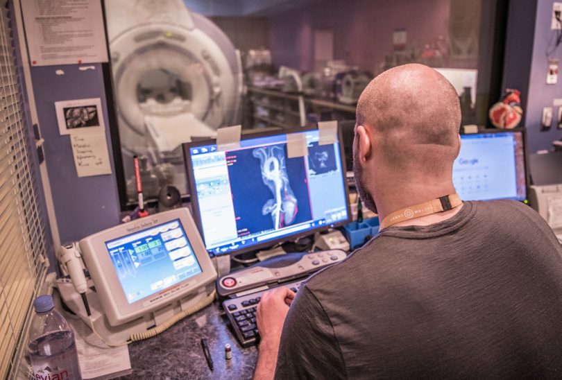
On a typical day at downtown Vancouver’s bustling St. Paul’s hospital, the Radiology department will perform an average of 20 chest computed tomography scans – better known as CT scans – on patients presenting with symptoms such as shortness of breath or chest pain.
This diagnostic test produces multiple pictures of the inside of the body, which can be reconfigured to produce three-dimensional images in order to detect various lung disorders such as benign and malignant tumors, pneumonia or cystic fibrosis.
False positives and negatives still common in diagnostic imaging
And while ever-advancing technology has resulted in diagnostic imaging that is clearer, faster and more accurate than ever before, detecting small lung malignancies still remains difficult; false positives and false negatives are not uncommon occurrences.
According to Dr. Jonathon Leipsic, Regional Head of the Department of Radiology for Providence Health Care and the Canada Research Chair in Advanced Cardiopulmonary Imaging, “There are so many images now that are generated, given the technological evolution of the scanners, that it can be a challenge to identify small lesions across so many images.”

Dr. Jonathon Leipsic, Regional Head of the Department of Radiology for Providence Health Care and Vancouver Coastal Health.
Currently, lung cancer is the leading cause of death from cancer in Canada, and early detection is vital in ensuring the best possible outcomes for patients.
So how can we improve the chances of early detection? Enter: artificial intelligence.
In a new study published last month in Nature Medicine, researchers from Google and leading medical centres in the US pitted a computer against expert radiologists to find out who could most correctly identify lung malignancies. The computer, armed with “deep learning” software that uses complex neural networks to identify patterns and uncover new knowledge, proved to be as good as radiologists at identifying lung cancer, and in some cases, performed even better.
From Nature Medicine: “When prior computed tomography imaging was not available, our model outperformed all six radiologists with absolute reductions of 11% in false positives and 5% in false negatives. Where prior computed tomography imaging was available, the model performance was on-par with the same radiologists.”
While the system is not yet ready for widespread adoption, the success of the initial findings is promising.
“This creates an opportunity to optimize the screening process via computer assistance and automation…(and) shows the potential for deep learning models to increase the accuracy, consistency and adoption of lung cancer screening worldwide,” say the study’s authors.
Artificial intelligence holds potential at Providence
Is this something radiologists here at Providence would welcome?
“If used thoughtfully, yes,” says Dr. Leipsic. “This could help facilitate improvements in care. Some artificial intelligence processes lend themselves to computer-aided assessment; lesion identification is certainly one.”
In fact, Dr. Leipsic’s team is already exploring the role that artificial intelligence might play in preventative medicine.
“My lab has been using AI to understand if there are imaging findings that can help us predict which patients are going to experience heart attacks and which patients are at risk of worsening of their emphysema,” says Dr. Leipsic. “There is great opportunity for artificial intelligence to reduce health care costs and advance our understanding of complex disease processes.”