A multi-year fundraising effort that garnered the support of many donors and ultimately raised $4.1 million of the $6.6 million project cost, has led to the St. Paul’s Hospital acquisition of a brand new 3T MRI (magnetic resonance imaging) machine.
The machine, operational at the hospital on June 14, 2021, is already having a tremendous impact on patient care.
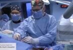
MRI is one of the gold standards in medical imaging. It lets practitioners see the body’s soft tissue, from ligaments and tendons, to its most complex regions such as the brain, spinal cord, and heart. It is widely used by cardiology, neurology, orthopedics, intensive care, and other areas to diagnose a range of complex pathologies and conditions.
More robust images of brain, other areas
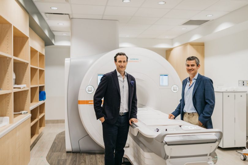
“This 3T MRI is not an overnight story; it’s really eight years in development,” says Dr. Jonathon Leipsic, director of Medical Imaging and regional department head at Vancouver Coastal Health and Providence Health Care (PHC). “We do thousands of MRIs every year. With this 3T magnet, we’ll be able to offer more robust imaging of the brain, image for many muscular-skeletal conditions, and really innovate in the cardiovascular and cardiopulmonary spaces.”
Faster scans, better images and single breath-holds
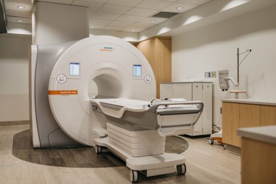
The new 3T MRI improves patient care on many levels. It provides faster scans and better image quality than the hospital’s two older machines, both of which are over 10 years old. With increased capacity, it is cutting down on wait times for patients. MRI is also very safe, and does not use radiation.
“As a tertiary referral centre, we look after some of the most complex cardiac patients in the province,” says Dr. Darra Murphy, head, Department of Radiology, PHC. “Some of our patients require long examinations that take two or three hours, involving multiple breath-holds, which can leave the patient quite exhausted. With this technology, we can do single breath-hold imaging that will really transform the quality of the imaging and the patient experience.”
3T MRI will have research applications into many diseases
Another major benefit is the 3T MRI’s application in research, into diseases like post-COVID syndrome, COPD, cystic fibrosis, asthma, and pulmonary fibrosis. The machine’s advanced technology enables clinicians and researchers to evaluate not only a patient’s lung disease progression, but also the impact of therapy and medications on their disease. Looking toward the future, St. Paul’s will be able to use the 3T MRI to transform the way we study lung, heart, and other diseases, with the promise of new breakthroughs in therapies and diagnostics.
“Our new 3T MRI will provide unique opportunities for research across all body systems but will really transform our capacity to investigate lung and heart conditions,” says Dr. Leipsic. “The polarized gas program we are setting up will be the first of its kind in Western Canada, providing a powerful tool to pair with the tremendous bench and clinical research emanating from our Centre for Heart Lung Innovation.”
“None of this promise and success would be possible without our donors,” says Dick Vollet, president and CEO of St. Paul’s Foundation. “Many people and organizations have stepped forward to make this life-changing 3T MRI a reality at St. Paul’s Hospital. In particular, we’d like to thank our four lead donors to this campaign: Cindy Liao; Sydney and Joanne Belzberg; Arnold and Anita Silber; and the Estate of William John Halchuk.”